
Why?
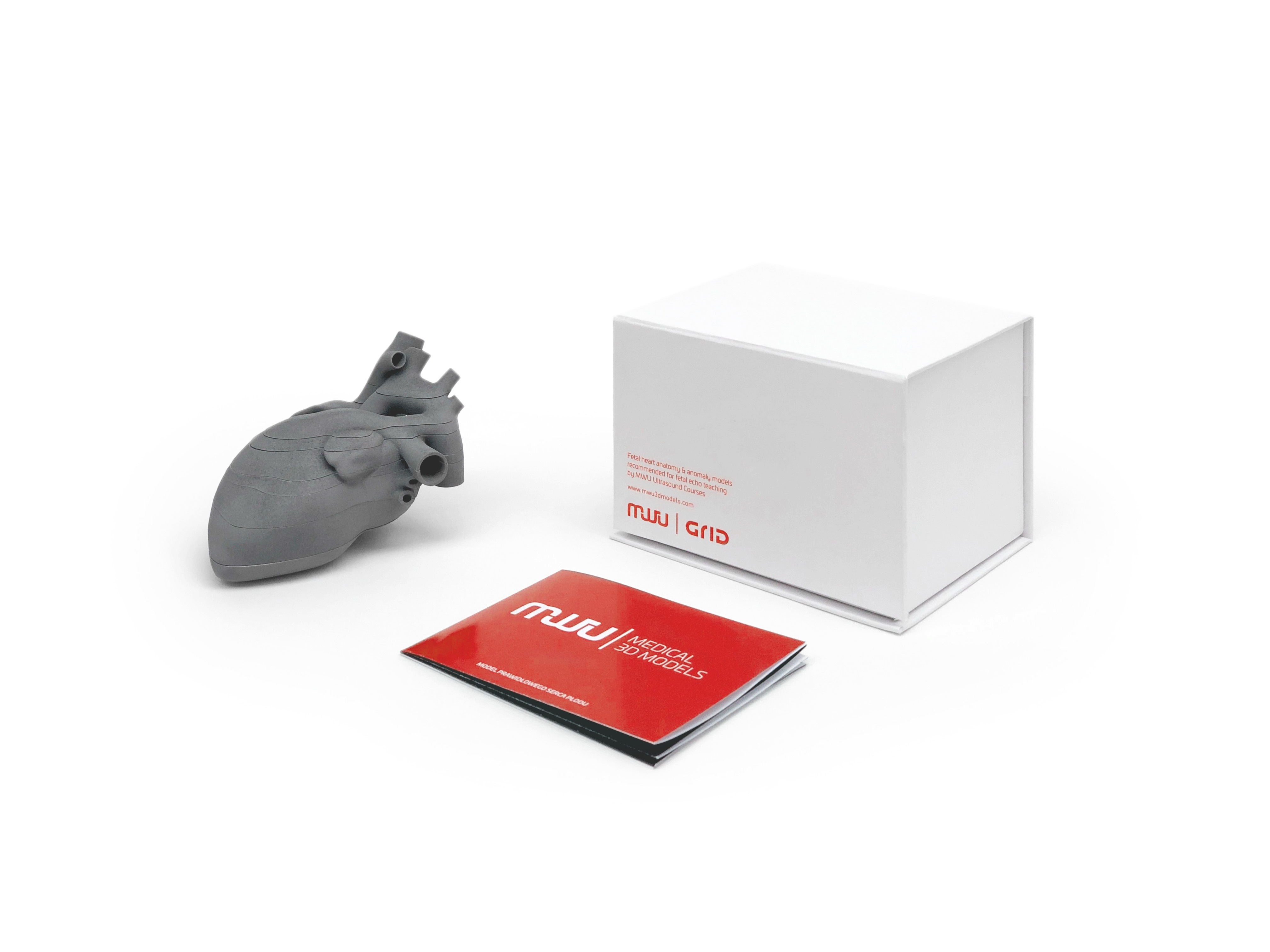
After years of teaching prenatal ultrasound we realized that sonographers, OB/GYN physicians and even fetal medicine specialists have difficulties in understanding three-dimensional aspects of the fetal heart. This element is crucial for better understanding of sonographic cardiac views and faster identification of Congenital Heart Defects (CHDs). We also noticed that commercially available cardiac models based on the adult heart lack the concept of arranging its elements in layers. Furthermore teaching prenatal echocardiography based on autopsy specimens has similar limitations including also the deficiency of appropriate tension, which is typical for the living fetal heart. Apart from teaching our models may be used as an important tool for counseling patients, which is far better understood by parents than traditionally used schemes and drawings.
For Whom?
After years of teaching prenatal ultrasound we realized that sonographers, OB/GYN physicians and even fetal medicine specialists have difficulties in understanding three-dimensional aspects of the fetal heart. This element is crucial for better understanding of sonographic cardiac views and faster identification of Congenital Heart Defects (CHDs). We also noticed that commercially available cardiac models based on the adult heart lack the concept of arranging its elements in layers. Furthermore teaching prenatal echocardiography based on autopsy specimens has similar limitations including also the deficiency of appropriate tension, which is typical for the living fetal heart. Apart from teaching our models may be used as an important tool for counseling patients, which is far better understood by parents than traditionally used schemes and drawings.
What is it?
Our 3D Fetal Heart Models are based on prenatal cardiac scans. They correspond with real ultrasound images, which are seen during a routine sweep through the fetal heart including four-chamber view, five-chamber view, three-vessel view, three-vessel and trachea view, and transverse aortic arch. Despite models presenting normal fetal hearts in transverse and sagittal views our project covers models of CHDs.
Recommendations
- MWU Courses (www.mwucourses.com )
- Dr. Fred Ushakov (Fetal Medicine Unit at UCLH, London UK; ISUOG Ambassador)
- Dr. Jacek Kolcz (consultant at the Department of Cardiac Surgery Ruprecht Karls University Heidelberg, Germany)
- Dr. Tomasz Roszkowski (senior consultant at Orlowski Distict Hospital in Warsaw, Centre of Postgraduate Medical Education in Warsaw )