Description
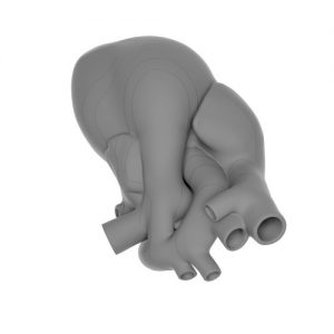
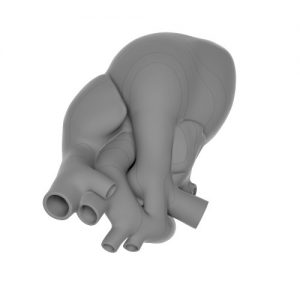
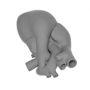
This is a 3D model presenting diagnostic features of ToF at 20 weeks of gestation in axial sections. Detection rates of this anomaly are still suboptimal. By better understanding of cardiac anatomy in ToF more cases will be diagnosed and especially the ones with ductal dependent circulation. The model shows all representative views including four-chamber view (4CV), five-chamber view (5CV), right outflow at the level of classical three-vessel view (3VV) wg Yoo, three-vessel and trachea view (3VTV), and transverse aortic arch view. Our model may be used on-line (with the presence of the patient) at the time of fetal cardiac evaluation in order to understand the mutual geometry of the segments and particular elements constituting abnormal cardiac anatomy of ToF. Our model is easy to use for the off-line learning (without the presence of the patient) by relating axial cardiac views demonstrated by means of video clips, still images or schematic drawings from textbooks or publications.