Description
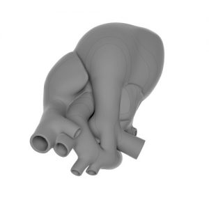
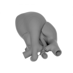
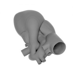
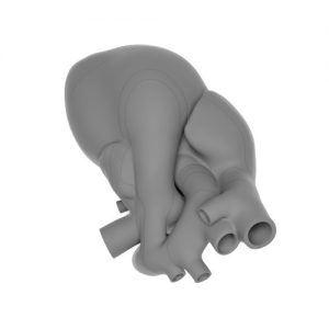
This is a 3D model presenting normal cardiac anatomy at 20 weeks of gestation in axial sections. It is a dextrocaria variant designed for the purpose of teaching. The reason is that levocardia variant may produce difficulties, when the fetal heart points with its axis to the right of the screen of the ultrasound machine. A dextrocardia variant model of the normal heart shows all representative views including four-chamber view (4CV), five-chamber view (5CV), right outflow at the level of classical three-vessel view (3VV) wg Yoo, three-vessel and trachea view (3VTV), and transverse aortic arch view. Our model may be used on-line (with the presence of the patient) at the time of fetal cardiac evaluation in order to understand the mutual geometry of the segments and particular elements constituting normal cardiac anatomy. This can be applied at any time, only if the fetal heart points with the cardiac axis to the right on the screen of the ultrasound scanner. Horizontal flip of the ultrasound image is required when the fetal heart points with its axis to the left on the screen. Our model is easy to use for the off-line learning (without the presence of the patient) by relating axial cardiac views demonstrated by means of video clips, still images or schematic drawings from textbooks or publications.